
A stage 1 pressure ulcer is reversible if identified within the first hours. After this period, the lesion progresses to involve the deep dermis and then the subcutaneous tissues, with a rapidly deteriorating healing prognosis. Recognizing a pressure ulcer at its onset relies on precise semiological criteria, often poorly understood outside specialized teams.
Beginning pressure ulcer on dark skin: a diagnosis often delayed
The NPUAP/EPUAP classification defines stage 1 as a non-blanchable redness on intact skin. On light skin, this sign is relatively easy to spot. On black or dark skin, the classic redness does not appear in the same way.
We then observe a darker, purplish or bluish area, sometimes shiny. Palpation reveals localized induration, abnormal warmth, or, conversely, coolness compared to adjacent tissues. Spontaneous pain in the sacral-gluteal area is an early warning signal, even in patients who do not easily verbalize.
This diagnostic delay on dark skin is documented: the absence of visible blanching leads to underestimating the lesion. Comparing a photo of an early pressure ulcer on different phototypes helps calibrate the clinical eye, especially for less experienced caregivers.
The practical recommendation: on dark skin, do not wait to see clear redness. Any change in color, texture, or local temperature in a pressure area should be treated as stage 1 until proven otherwise.

Semiology of stage 1 at the gluteal area: distinguishing the ulcer from a simple erythema
The confusion between a reactive erythema (related to moisture, maceration, or friction) and an early ulcer is common. The distinction relies on a simple test: the vitropression.
Press firmly with a finger or a transparent disc on the red area for three seconds, then release. If the skin whitens under pressure and then turns red again, it is a reactive erythema. If the redness persists without blanching, it is a stage 1 ulcer.
Other clues guide the diagnosis:
- A localized pain disproportionate to the skin appearance, reported by the patient or noticed during mobilizations
- A localized edema at the sacrum or ischium, perceptible on comparative palpation
- A change in the firmness of the subcutaneous tissue: a harder or conversely softer area than the surrounding tissues
These signs precede ulceration. A stage 1 pressure ulcer shows no open wound. This is precisely what makes it difficult to detect for untrained caregivers, who often associate “ulcer” with “visible wound.”
Recommended frequency of skin assessment
For a bedridden or wheelchair-bound patient, we recommend a visual and tactile inspection of the gluteal pressure areas at every position change. The 2001 consensus conference (ANAES, now HAS) emphasizes this systematic monitoring, particularly at the sacrum and heels, which account for the vast majority of cases.
Pressure relief on the gluteal area: the priority action from the first signs
In the face of an early pressure ulcer, the most effective reflex is neither a local treatment nor a topical product. Immediate relief of pressure on the affected area determines the reversibility of the lesion.
Specifically, this means:
- Removing all pressure from the affected area by changing the patient’s position (lateral decubitus if the ulcer is sacral, sitting if the ischium is involved, and vice versa)
- Alternating positions every two to three hours at most, including at night, avoiding resting the patient on the affected area
- Using an appropriate pressure redistribution support (alternating air mattress, positioning cushion), not a simple standard foam cushion that collapses quickly
- Reducing shear forces: do not raise the head of the bed beyond 30 degrees, limit the patient’s sliding on the sheets
Friction worsens the lesion. During repositioning, lifting the patient rather than dragging them on the sheet reduces skin shear. Using a slide sheet facilitates this maneuver.
Risk factors often overlooked outside the hospital setting
At-risk individuals are not only bedridden patients in institutions. Prolonged sitting in a wheelchair, but also on a standard seat (sedentary work, long-distance transport) generates sustained pressure on the ischium and sacrum.
Urinary or fecal incontinence, protein malnutrition, and dehydration weaken the skin and accelerate the progression of an early lesion. A degraded nutritional status significantly prolongs the healing time, even at stage 1.
Local care at stage 1: what to do and what to avoid
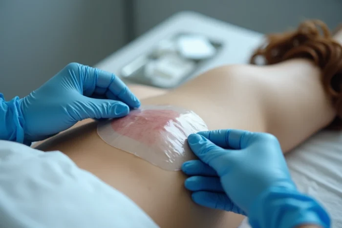
At stage 1, the skin is intact. No dressing is necessary, and massaging the reddened area is strictly contraindicated: massage worsens deep tissue lesions by increasing subcutaneous shear.
The course of action is limited to keeping the skin clean and dry, without rubbing. Use a neutral pH soap, pat dry. Moisturizing creams can be applied to the surrounding healthy areas to maintain skin elasticity, but not on the injured area itself.
Skin protectants like semi-permeable films or thin hydrocolloids are sometimes used in secondary prevention on at-risk areas, but their indication at stage 1 remains debated among teams.
When to alert a healthcare professional
If the redness does not regress within 24 to 48 hours despite the removal of pressure, the lesion is likely progressing to stage 2. The appearance of a blister (serous or hemorrhagic) or superficial epidermal loss requires prompt consultation, ideally with a nurse trained in chronic wounds or a physician.
Early management of a pressure ulcer relies on a simple action (relief) and a trained eye (systematic vitropression). For at-risk patients, regular skin assessment of pressure areas remains the most effective preventive measure, well ahead of any technological device.